Behind the Lens
Behind the Lens
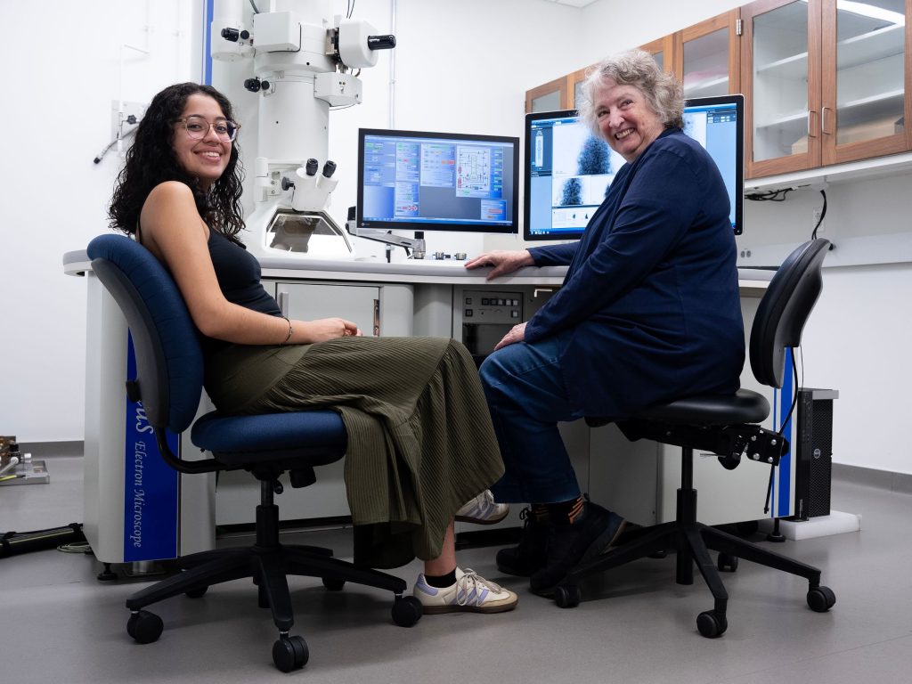
For more than four decades, Nancy Piatczyc has been helping Williams students and faculty see the unseeable—at least to the naked eye. As the electron microscopy technician, Piatczyc is the steward of powerful microscopes in the Electron Microscopy Lab, where she maintains the equipment, shows students how to use it and provides technical support when needed.
Students like Andrea Bustos ’26, a geosciences major from Miami, Fla., (pictured with Piatczyc above) use the lab’s microscopes—a rare opportunity at a small liberal arts college—for their senior thesis research. Piatczyc, who earned a B.S. in biology from Tufts University and worked in labs and hospitals before coming to Williams, says, “It’s unusual for undergrads to get to use the electron microscopes, as most colleges and universities only allow graduate students to use this equipment.” She’s quick to note that Williams students across the sciences and other disciplines can also access these state-of-the-art instruments.
Located on the lower level of the Hopper Science Building, the Electron Microscopy Lab houses a scanning electron microscope (SEM), a transmission electron microscope (TEM), a confocal microscope and an atomic force microscope. In addition to showing students how to use them, Piatczyc also demonstrates how to prepare specimens—bacteria, fossils, bugs, tiny plant particles and other matter—for imaging.
Recent research involving the electron microscopes has included geology students looking at the morphology and chemistry of thin sections of rock on the SEM. Labs for courses in geology, chemistry and biology used that microscope, too, to examine specimen samples. Chemistry labs have also used the SEM to look at nanoparticles in the sub-micron range. And biology students have conducted a study of fish spinal cords and the fine structure of nerves using the TEM. When not in use during regular semesters, the microscopes are used during Winter Study and summer programs.
Piatczyc points out that electron microscopes are in demand outside the sciences, too. For example, at the Williamstown Conservation Center, located on the grounds of the nearby Clark Art Institute, “Conservators can take a tiny speck from a painting or material from a sculpture and get an elemental analysis,” she says. “They’re often looking to see what the original painting is made of, either for restoration or to determine if the work is a forgery.”
After 42 years at Williams, Piatczyc will retire in August. A bittersweet occasion, she says that she’ll miss the students and faculty most of all. “They are really fantastic and so easy to work with,” she says with a smile. But the microscopes themselves, and the incredible images and understanding they bring to light, is something she says never gets old. “Sometimes when I’m waiting for students to come, I’ll image various things like plant leaves, just to see the pollen,” she says. “It’s so much fun.”